Spine – Anterior artificial discs & cages + Posterior surgery options
I have done many anterior cervical and lumbar reconstructions over several years. My patients receive appropriate (pathology- and anatomy-tailored) combinations of state-of-the-art artificial discs and cages.
Whenever feasible, I prefer anterior reconstruction to posterior reconstruction because I believe anteriorly instrumented patients (where complete discectomy can be done and large-footprint prosthetics can be inserted, and where there is at least an option for natural movement when desired/needed) do better than the traditional posterior fusion patients (lots of tissue dissection, typically small and unanchored ‘bullet’ cages, incomplete discectomies, no good natural movement option). However, there is certainly a good place for the latter depending on pathology, body habitus, medical factors – i.e., with careful patient selection.
Welcome to Dr Khurana’s Spinal Instrumentation page.
To begin with, CLICK HERE for a great video to show you how a broken spine is fixed; it’s the best I can do! (noting I could not use an artificial disc in that particular lady’s situation, given the profound ‘slip’ she had as you’ll see).
Return to this page via your back arrow – more videos and information below.
Click the following links for brief and informative step-by-step videos or blogs on Dr Khurana’s “total disc arthroplasty (TDR)” aka “spinal arthroplasty” surgeries:
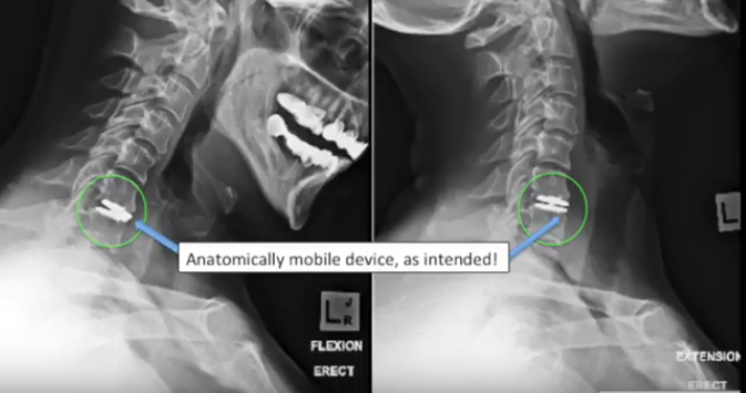
Video: ARTIFICIAL MOBILE CERVICAL DISC SURGERY
Video: ARTIFICIAL MOBILE LUMBAR DISC SURGERY
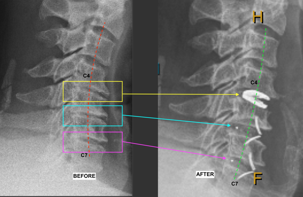
CLICK HERE to read more about “a thing of beauty” – restoring the collapsed, kyphotic neck with longstanding axial neck pain
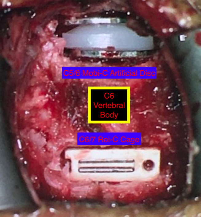
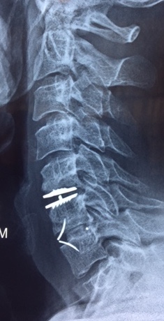
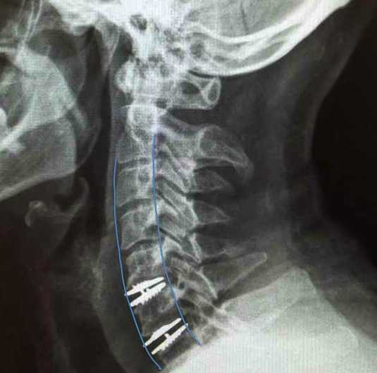
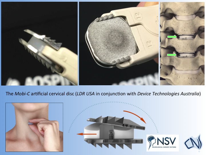
Above images (click each or use your zoom to enlarge): The latest in “minimal footprint” hybrid cervical disc replacement technology (Left, intraop.; Right, three months post-op.) — no plates, no screws, no cervical collar; the upper artificial disc inserted to preserve mobility and reduce wear-and-tear at adjacent segments; operation carried out by Dr Khurana at East Sydney Private Hospital (Intraoperative view; Mobi-C, ROI-C; courtesy Device Technologies/LDR). A beautiful structural and functional result.
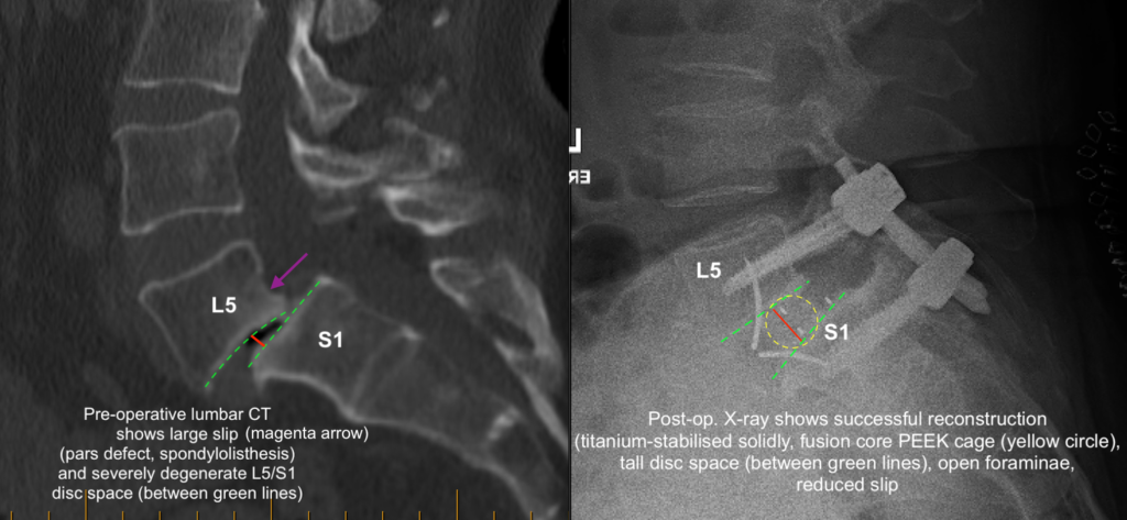
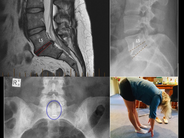
Below images (click each or use your zoom to enlarge): The latest in “minimal footprint” lumbar disc replacement technology (Images a&b: Pre-op. CT & SPECT showing severe degeneration at L4/5 and L5/S1 with “deflated and worn tyre” disc spaces and a slip at L5/S1; Image c: The ROI-A cage. Image d: Post-op. CT showing beautiful restoration of disc heights and reduction of slip via insertion of ROI-A cages through a small abdominal incision – no plates, no screws, no hip graft, just minimally invasive techniques and technologies in action; courtesy Device Tech./LDR).
a.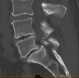 b.
b.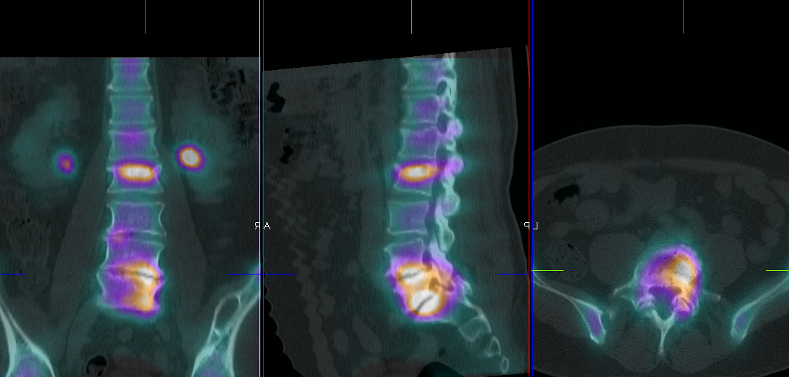 c.
c.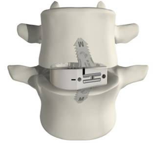 d.
d.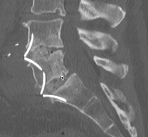
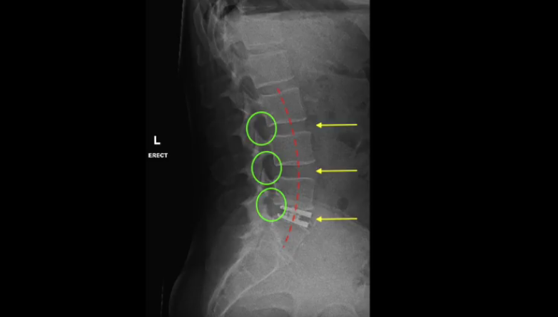
RANGE OF SPINAL MOTION (below collage) a year after L5/S1 ALIF using ROI-A cage system via anterior (“bikini-line”) minimally invasive approach. Read more on this patient’s case by CLICKING HERE.
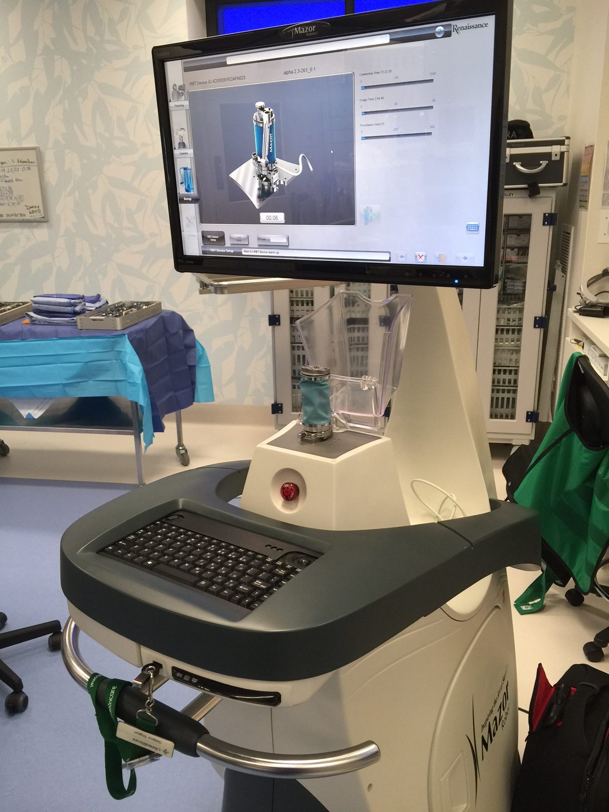
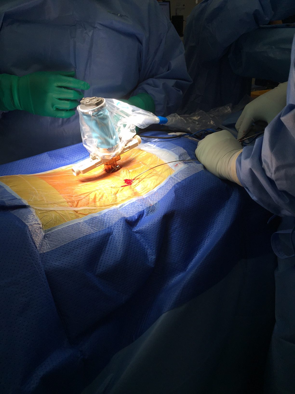
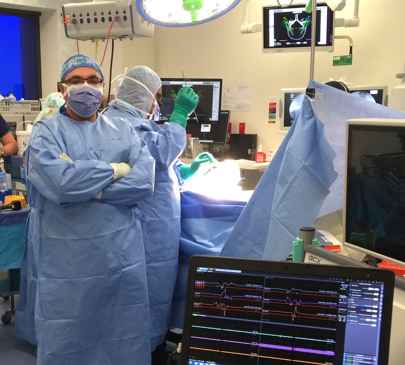
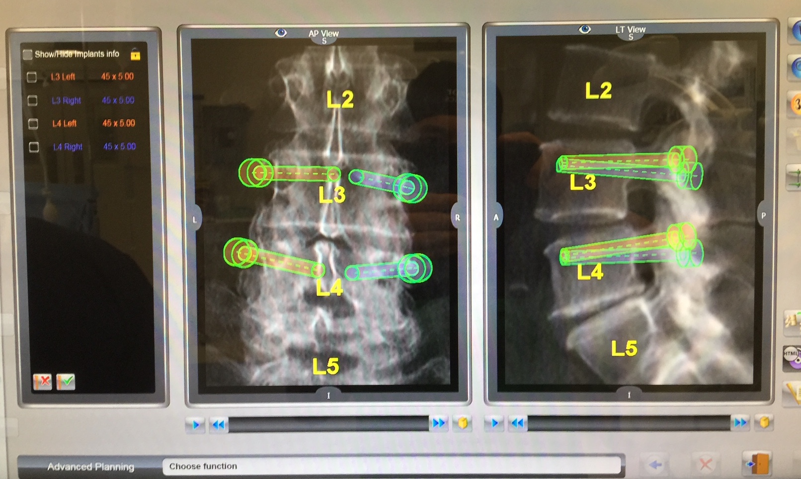
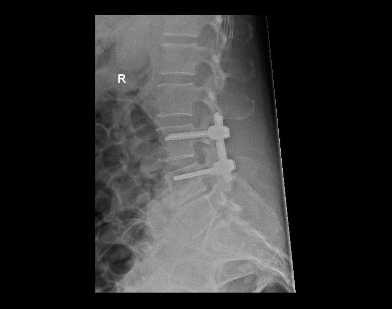
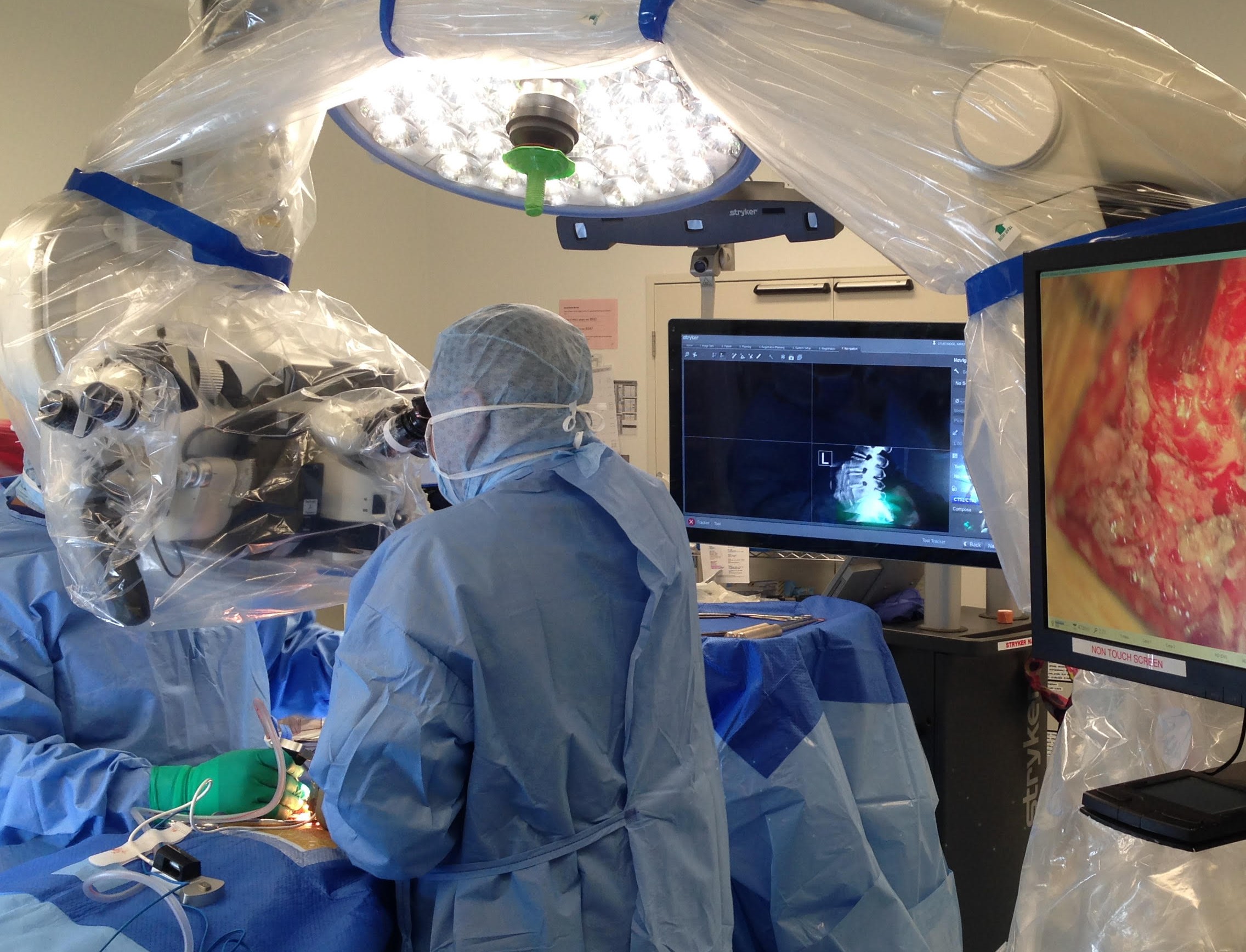
ROBOTIC SPINAL SURGERY has also been introduced into our Practice. Immediately below, see images of the Mazor Robot with Renaissance Guidance System in action at the Epworth Hospital, with real-time neuromonitoring of the patient during the procedure. The Robot has been introduced because it has been internationally tested and reported to be: Intuitive. Quick. Precise. Safe.
[With thanks to: Epworth Hospital, Life Healthcare, Mazor, Neuro-monitoring Services Australia]
Images shown here are with the permission of Dr Khurana’s patients, for educational purposes.
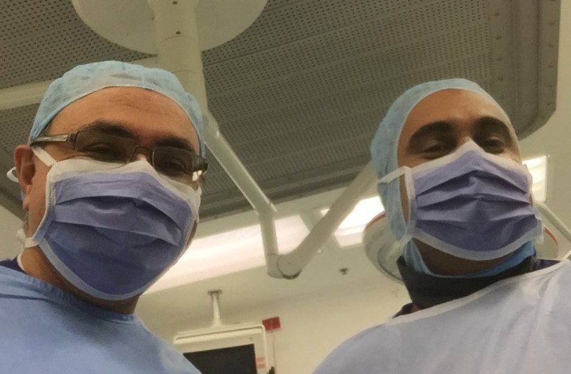
- Neurosurgeons Mr Vini Khurana & Mr Ron Jithoo.
Shown on this page are minimal-footprint artificial spinal discs and interbody cages, and Robot-navigated screws… ‘state-of-the-art’ technologies and minimally invasive options and techniques designed and tailored to minimise disturbance to your natural tissues and maximise your functional outcome.
-
- Image 1
-
- Image 2
-
- Image 3
-
- Image 4
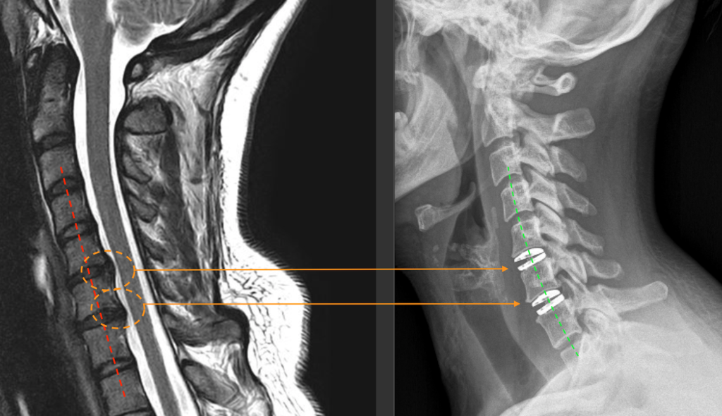
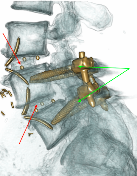
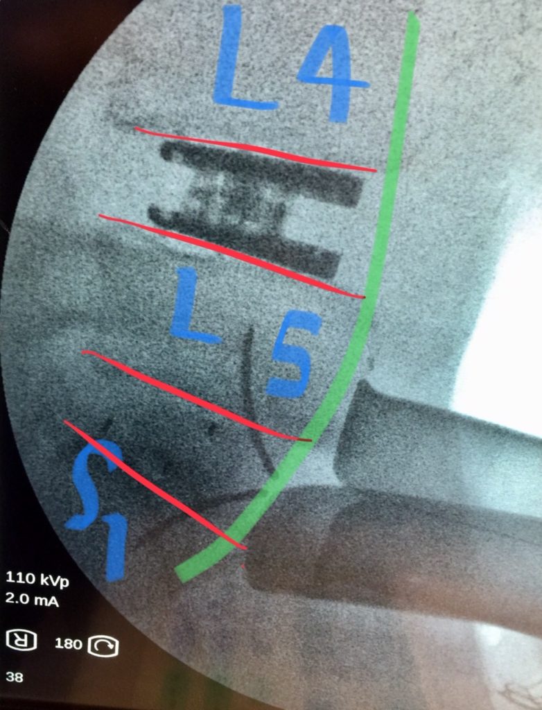
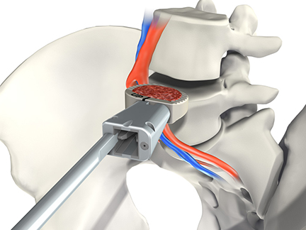
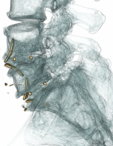
Image 8 (above): The ROI-A lumbar cage (filled with a natural bone tissue scaffold-matrix) is placed using a minimally invasive surgical approach via a small abdominal incision (aiming to facilitate a quicker and smoother recovery). No screws, no plates, no hip graft. Typically, even our 2-level ROI-A patients are up and walking the day after surgery.
CLICK HERE for more images of advanced spinal instrumentation surgery