A ‘thing of beauty’ – State-of-the-art anterior hybrid cervical reconstruction: Before vs. After – Artificial disc, cage prosthetics
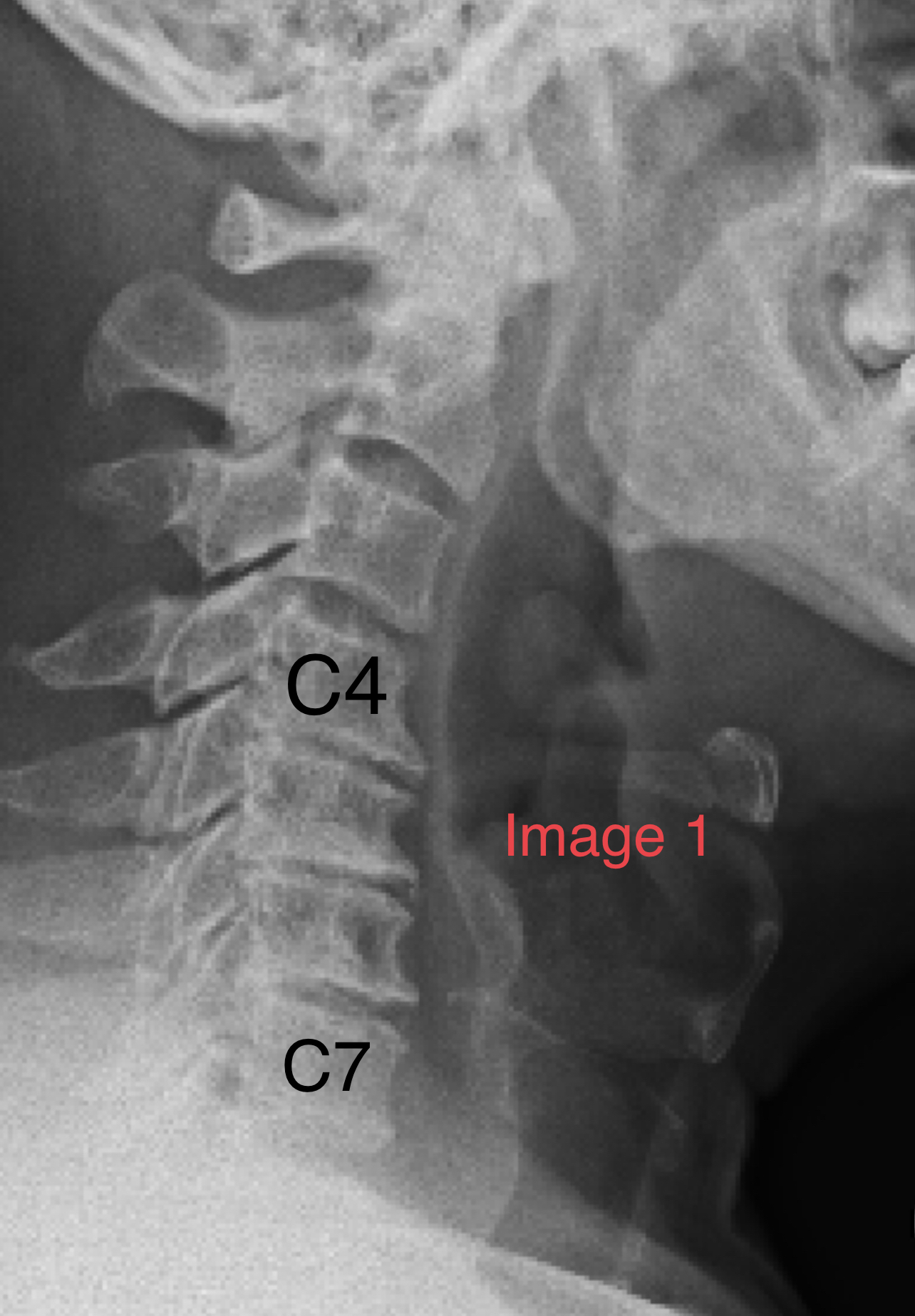
Image 1 shows the state of this man’s neck on a lateral X-ray. Mid-60s, with over 40 years of axial neck pain.
Pre-operatively, I felt his neck to be relatively “collapsed and kyphotic” especially at C4/5, C5/6 and C6/7. The normal lordosis (slight ‘backward bending’ or ‘anteriorly convex’ curvature) of the cervical spine is also quite ‘reversed’ here.
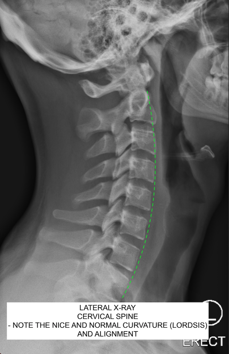
Just to compare, this is what a younger person’s healthy neck looks like with normal lordosis and nice, tall, cushion-like disc spaces:
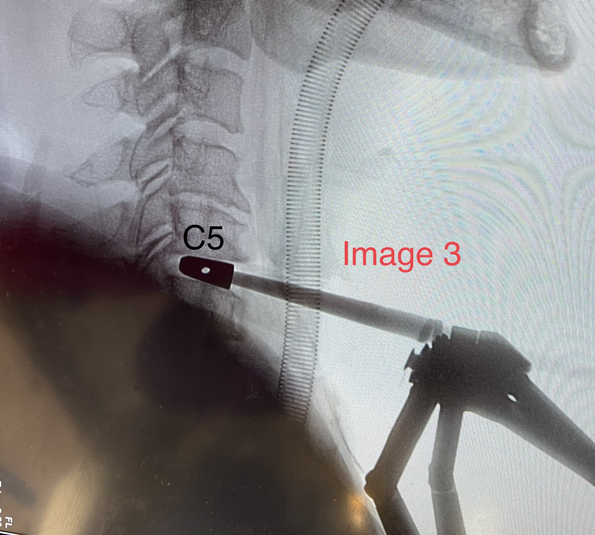
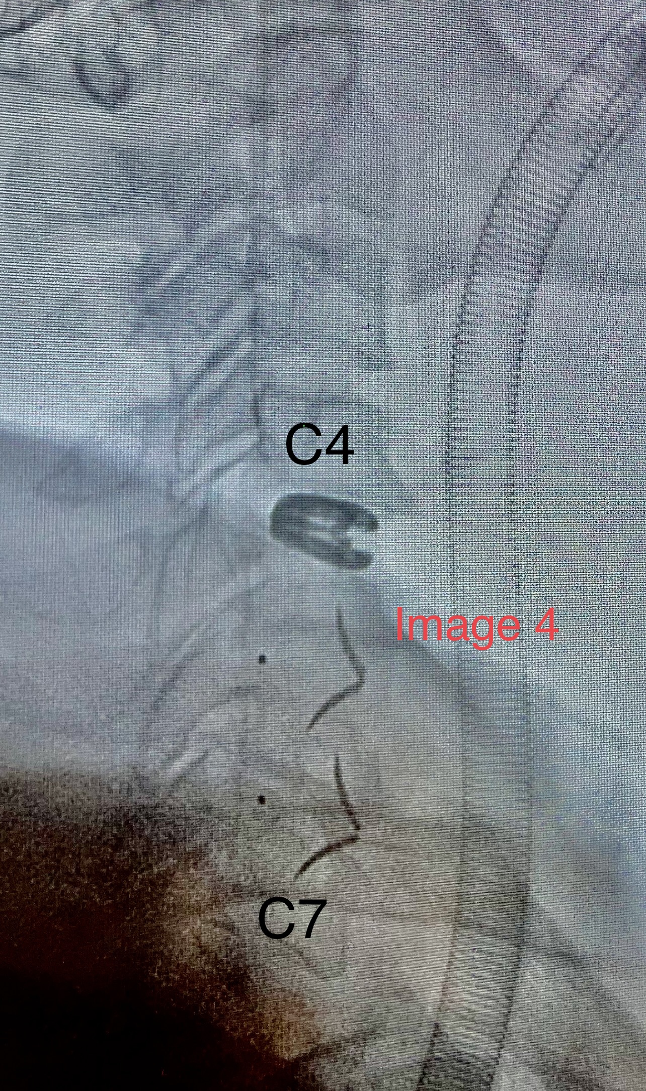
Intraoperatively, I felt that his C4/5, C5/6, C6/7 disc spaces had probably not seen any movement for many years, as they were osteophytic externally and part-ossified internally, and completely worn out with little endplate cartilage remnants. I removed each of the worn discs sequentially, and treated the cord compression he had at C5/6 (normal cord shown in Image 2 after decompression at C5/6).

Prosthetics were chosen one at a time for size, shape, best fit and functionality (structural support, reshaping and/or mobility).
These are my preferred prosthetics for this operation, and I believe they are the very best out there. I chose to put in a mobile artificial disc in C4/5, and cages at C5/6 and C6/7 (“hybrid prosthetic construct” or just “hybrid“). These lower two spaces were not suitable for the disc owing to a lack of decent anchoring tissue, and lack of a dome-shaped superior endplate, and I prefer not to place an artificial disc if there is cord compression (C5/6 in this man).
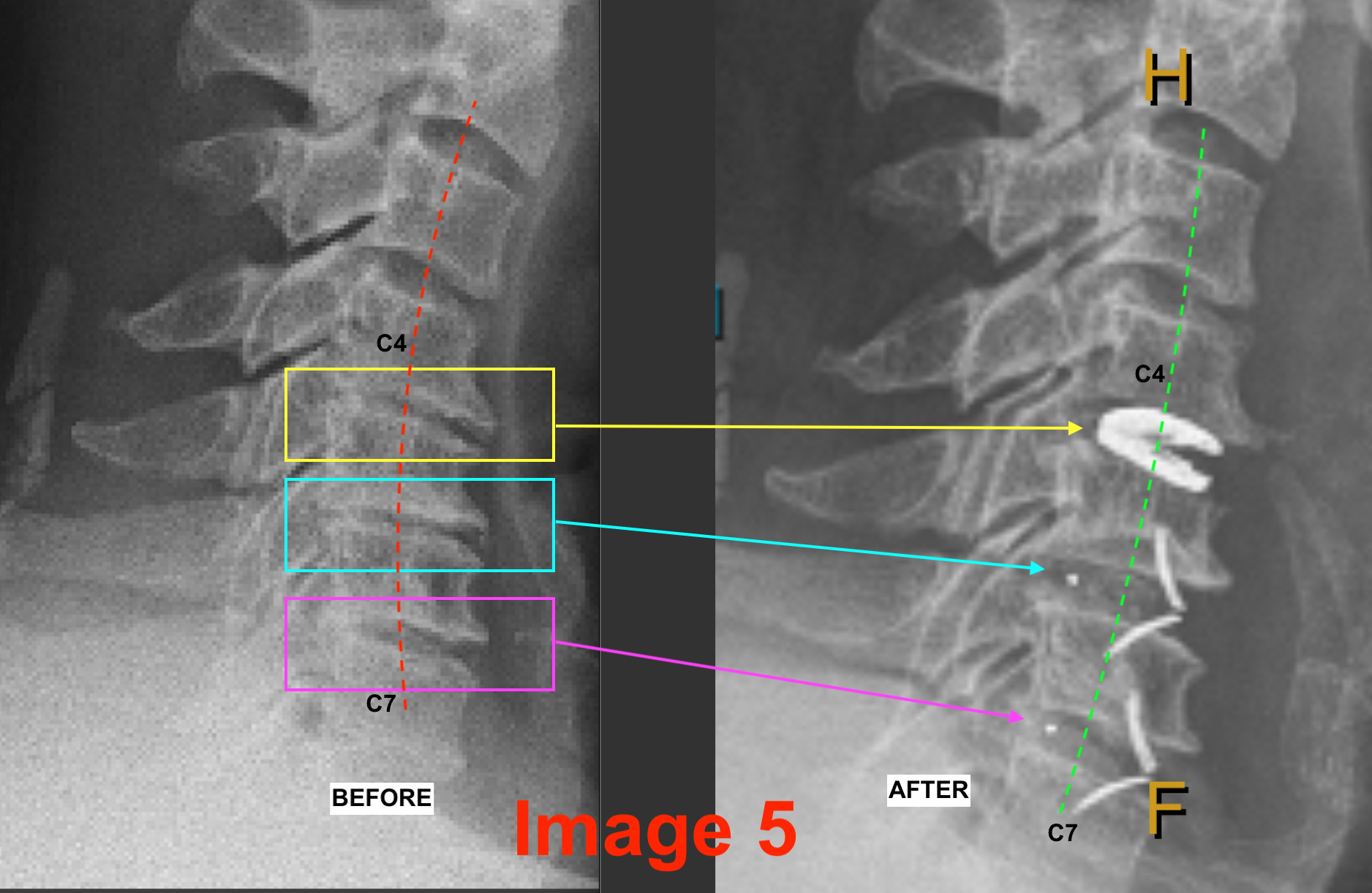
Below is the “before” versus “after” lateral X-ray shot. I think it’s a thing of beauty. Look what we started with anatomically, and look where we ended up prosthetically (structurally/functionally). The kyphosed neck (red dashed line) is now better lordosed (green dashed line), and the previously collapsed disc spaces (boxes) have nice heights and shapes (arrows) at C4/5, C5/6 and C6/7. This is us as reconstructive surgeons trying to restore cervical lordosis (lordotic restoration) when the cervical spine is alordotic (straight) or early reverse-lordotic (curved the “other way”, like in this man; see image 1). It’s a small miracle to have such amazing implants available to my patients in this day and age. Sleek, no bulky plates, no screws.