“Hybrid” anterior cervical and lumbar reconstructions – spinal resculpting, stability & mobility
Minimally invasive “hybrid” cervical and lumbar reconstructions can be accomplished using a combination of one or more mobile artificial discs (total disc replacement/TDR) and sleek cages (anterior interbody fusion). These can be done through small incisions in the neck and abdomen. By 3 months post-operatively, my “hybrid” patients quite uniformly report substantially improved symptoms and mobility following these surgeries.
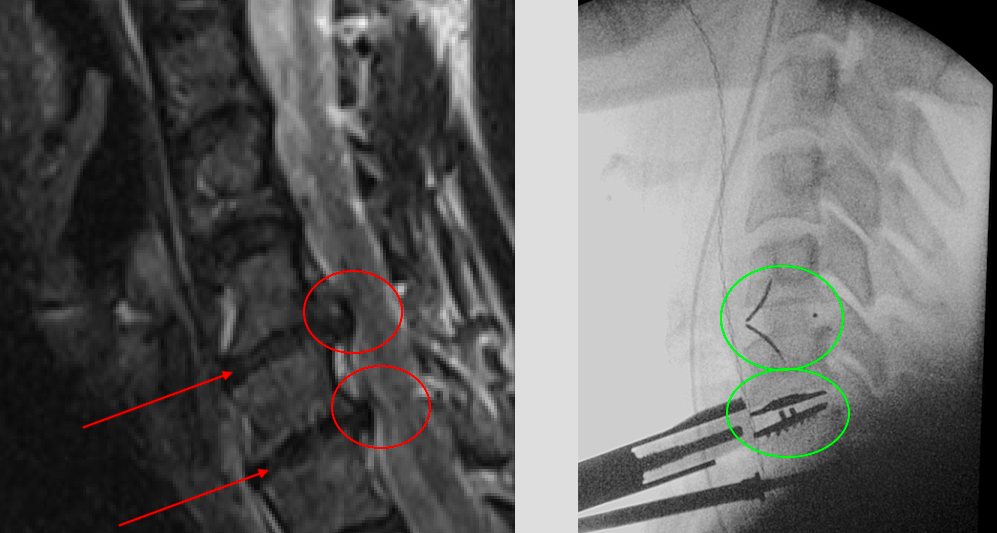
This patient (see below) in her 40s had symptomatic cervical cord compression from two really worn, collapsed and herniated discs (lower left; red arrows and ovals). The discs were removed via a small lower front neck incision, and a hybrid reconstruction carried out with the intra-operative x-ray of the construct shown (lower right; green ovals). A ROI-C cage was put in by me at C5-6 and a Mobi-C disc put in at C6-7. Her natural cervical curve (lordosis) has been improved by the surgery, in addition to the decompression of her cervical spinal cord and preservation of her neck’s natural mobility and strength. The materials should last her lifetime; they are a combination of titanium and re-inforced plastics, with fusion biomaterials in the core of the cage at C5-6.
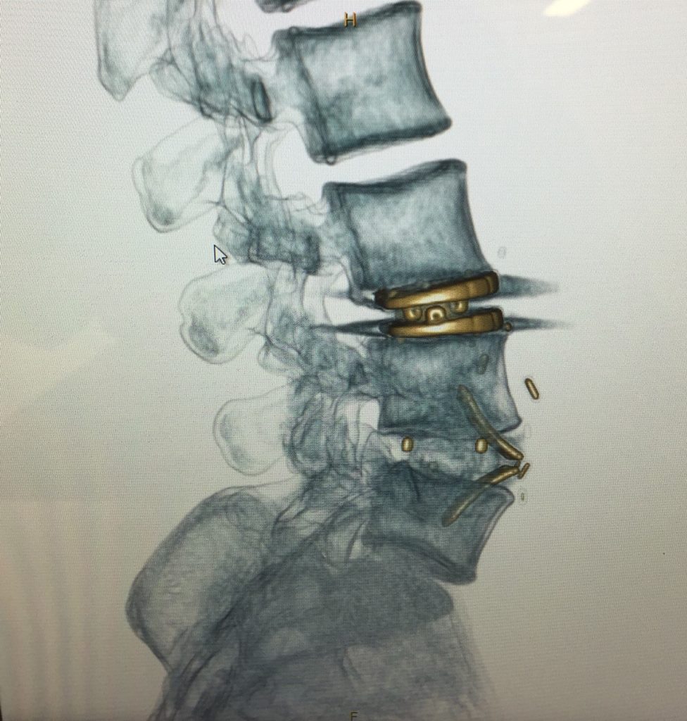
Similar “lumbar hybrid” surgery can be carried out where appropriate in the lumbar spine (CT “3D” reconstruction image shown below, following ESP-LP artificial disc insertion by me at L3-4 and ROI-A cage insertion at L4-5). This surgery was carried out via a small anterior abdominal incision made by the expert vascular and tranplant surgeon, Miss Sharmila Balanathan, who I operate with for ALIF/LTDR procedures.
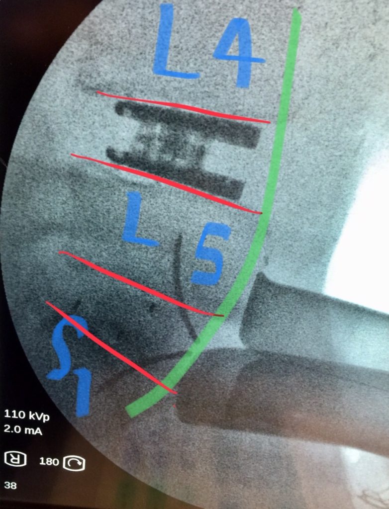
Restoration of shape (lordosis; green line), disc height (between adjacent red lines), structure (whole construct) and stability (L5 and S1 titanium “gull-wing” anchors and cage central fusion core), while releasing pressure on compressed local nerves, is all possible through anterior mininally invasive reconstruction such as this (image below, L4/5 artificial ESP-LP disc; L5/S1 ROI-A cage)
For more information, take a look at CNS Neurosurgery’s other blogs or visit our Resources page.
< Back to blog