Normal versus abnormal lumbar anatomy
This set of 4 images below is illustrative of normal lumbar anatomy versus abnormal lumbar anatomy. The images can be used in conjunction with information that CNS Neurosurgery has provided on our Spondylosis page.
Image below: Normal lumbar anatomy as seen on a sagittal T2 MRI.
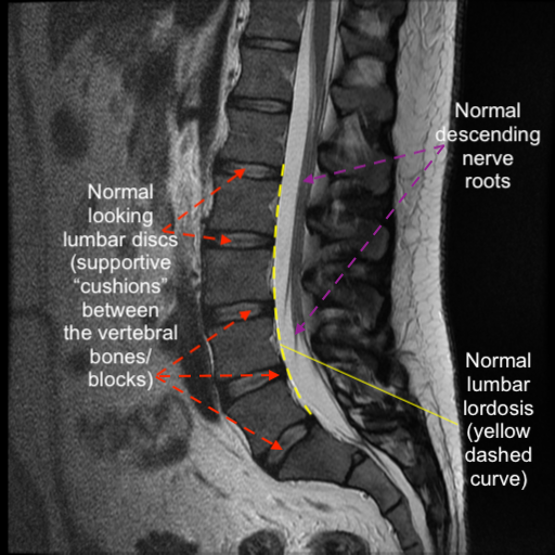
Image below: Normal lumbar anatomy as seen on an axial T2 MRI.
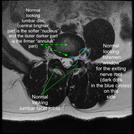
Image below: Abnormal lumbar anatomy as seen on a sagittal T2 MRI.
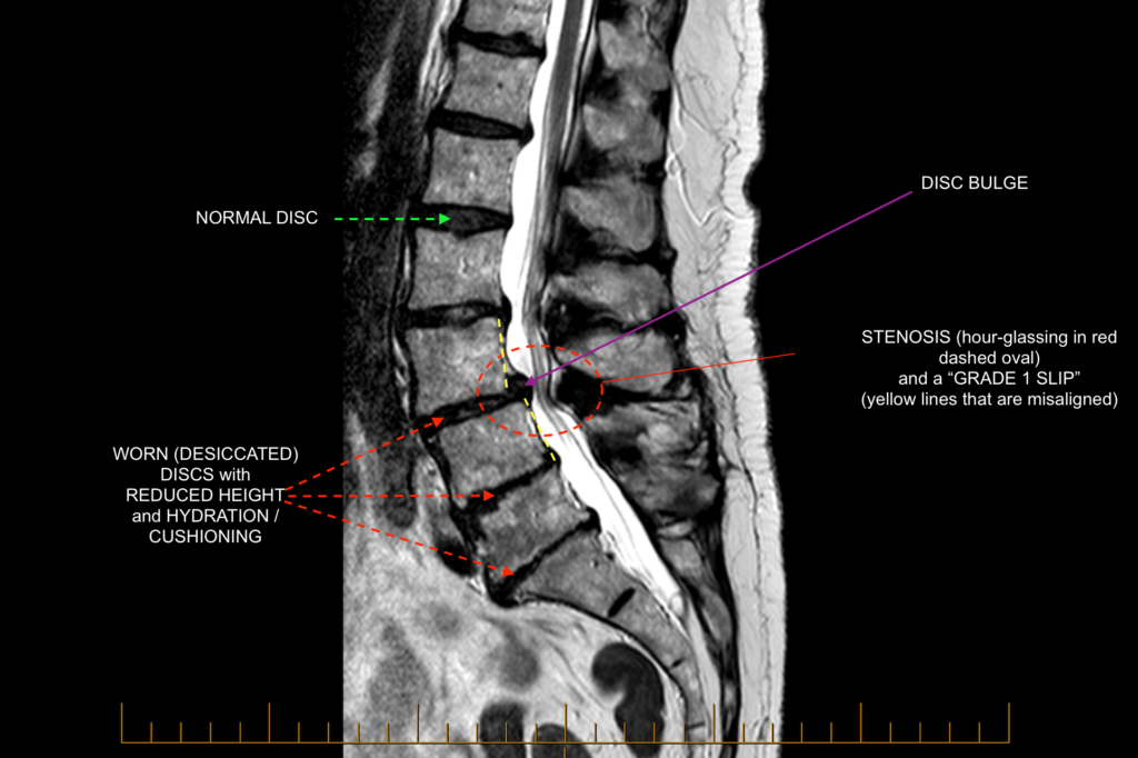
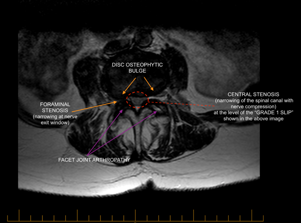
Image below: Abnormal lumbar anatomy as seen on an axial T2 MRI.