Radiological follow-up of some of my anterior lumbar prosthetic patients: ROI-A fusion cages and LP-ESP artificial discs at 1 to 6 years post-insertion
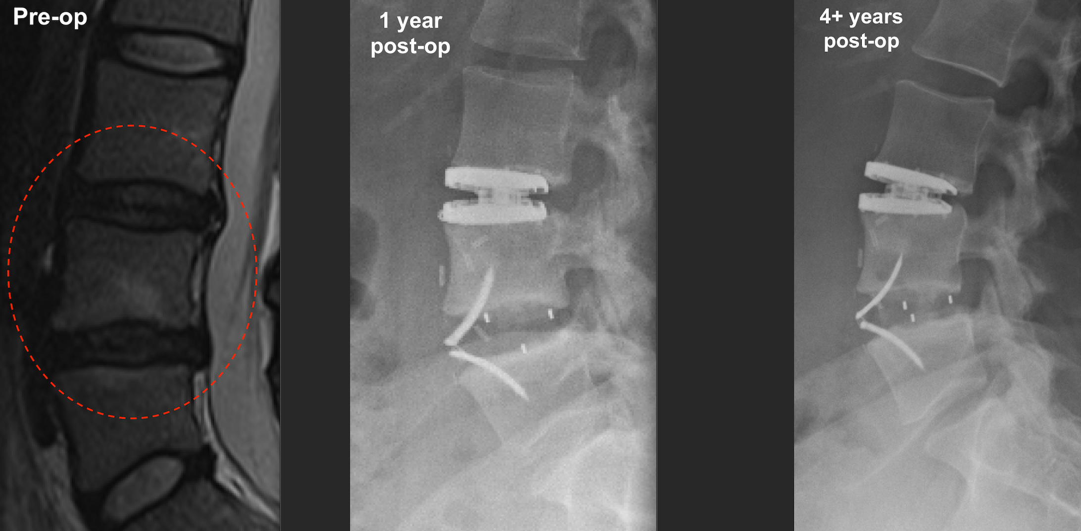
The anterior lumbar “hybrid” surgery patient’s images below involved replacement of her worn, damaged and symptomatic L3/4 and L4/5 discs (left panel, Pre-op) with an LP-ESP artificial disc at L3/4 and a ROI-A fusion cage at L4/5. The centre panel shows the prosthetics at 1 year post-op and the right panel shows them at 4+ years post-op. The cage shows evidence of progressive central fusion as expected. The operated disc spaces remain tall and satisfactory and there is preservation of lumbar lordosis as intended. There is no obvious lucency around the prosthetic components to indicate any loosening, and there is no displacement during the follow-up. They look well integrated, as I had hoped. This is at 4+ years of post-operative clinicoradiological follow-up.
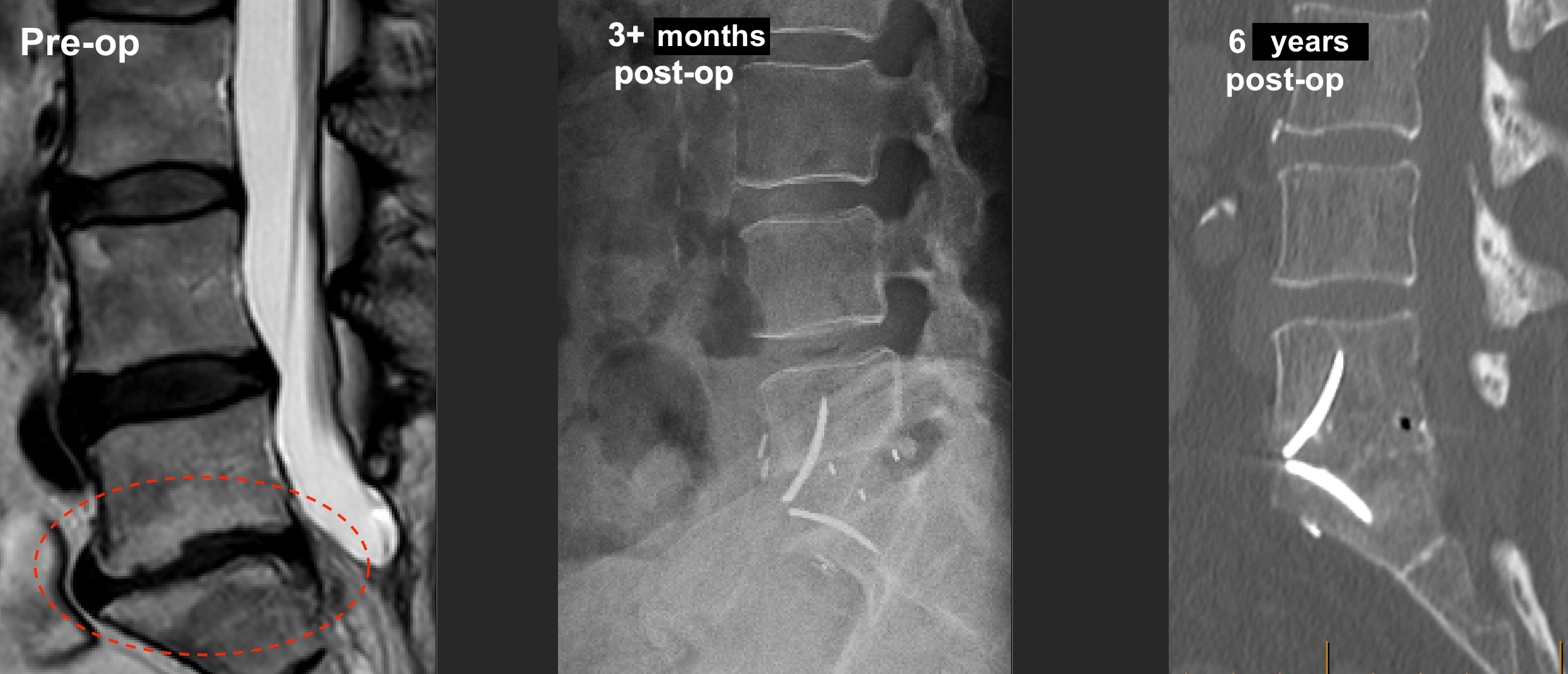
The anterior lumbar interbody fusion (ALIF) surgery patient’s images below involved replacement of the severely worn, damaged/desiccated and symptomatic L5/S1 disc (left panel, Pre-op) with a ROI-A fusion cage. The centre panel shows the prosthetic at 3-4 months post-op and the right panel shows it at 6 years post-op. The cage shows evidence of progressive central fusion as expected. The operated disc space remains tall and satisfactory and there is preservation of lumbar lordosis as intended. There is no obvious lucency around the prosthetic components to indicate any loosening, and there is no displacement during the follow-up. The cage looks well integrated, as I had hoped. This is at 6 years of post-operative clinicoradiological follow-up.
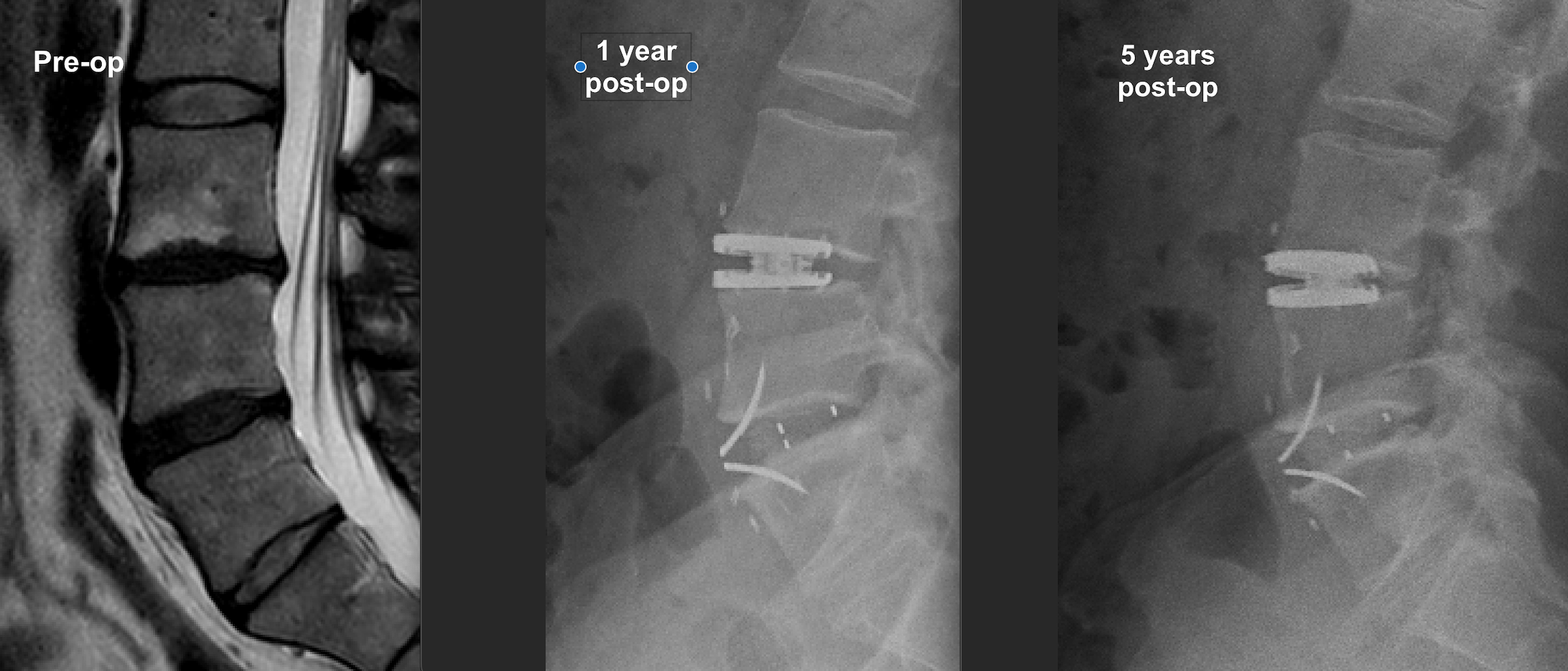
The anterior lumbar “hybrid” surgery patient’s images below involved replacement of his worn, damaged and symptomatic L3/4 and L4/5 discs (left panel, Pre-op) with an LP-ESP artificial disc at L3/4 and a ROI-A fusion cage at L4/5. The centre panel shows the prosthetics at 1 year post-op and the right panel shows them at 5 years post-op. The cage shows evidence of progressive central fusion as expected. Again, the operated disc spaces remain tall and satisfactory and there is preservation of lumbar lordosis as intended. There is also no obvious lucency around the prosthetic components to indicate any loosening, and there is no displacement during the follow-up. They look well integrated, as I had hoped. This is at 5 years of post-operative clinicoradiological follow-up.