Visual example comparing the resolution of 3T MRI vs. (standard) 1.5T MRI in spinal imaging
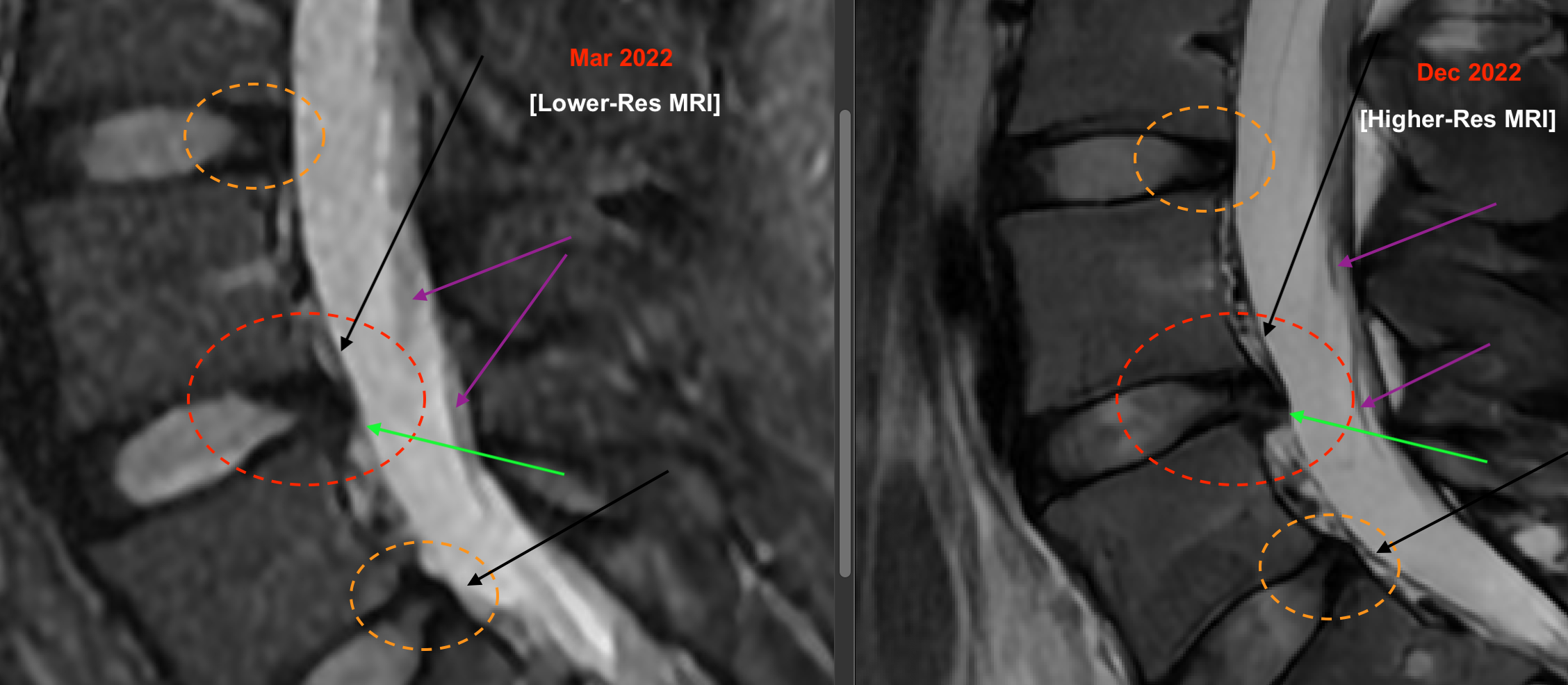
The same patient underwent a standard 1.5T (T=Tesla) MRI of the lumbar spine in early 2022 (left panel, above), and then a 3T MRI of the lumbar spine in late 2022 (right panel, above). You can see the left image (1.5T magnetic field strength) is of much lower resolution than the right image (3T magnetic field strength). Compare the regions of posterior disc desiccation (yellow dashed ovals), disc herniation (red dashed ovals), posterior longitudinal ligament (PLL; tips of black arrows), and descending cauda equina nerve roots (tips of magenta arrows). A nice example, I think, of potential resolution and diagnostic improvement readily achievable these days. There is no radiation involved in this modality, just a magnetic field, but patients should check with their doctors and imaging providers if any current or planned prosthetic implants in them are compatible with MRI, and with 3T MRI.
< Back to blog