Artificial disc + cage anchor & fusion points for spinal biointegration: “Hybrid” prosthetic construct shown here
My patients often ask about which parts of the prosthetics that I use during cervical and lumbar anterior reconstruction (i.e., my cages and artificial discs) are meant to “fuse” and which parts are “not meant to fuse“, and how are they “anchored in“?
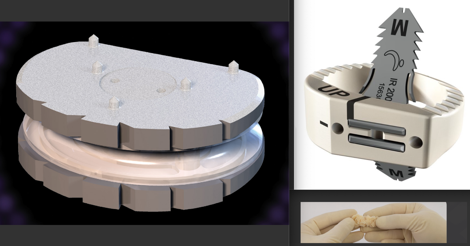
Image 1 below shows my preferred prosthetics: An FHO ESP artificial disc (coated titanium plates with a silicone gel + polycarbonate urethane/PCU core) on the left; a LDR ROI cage (ultra-strong PEEK plastic with titanium gull-wing anchors) with Boost Ultra fibers (a bone-forming ‘putty’ for its core) on the right (BTW, these French designed and manufactured discs (FHO company) and cages (LDR company) are magnificent, and I’ve been successfully implanting them for years).
So, here’s an illustrated example from a recent anterior hybrid lumbar spinal patient of mine. Note that the same applies to the cervical spine, as I use the same types of state-of-the-art discs and cages at both locations.
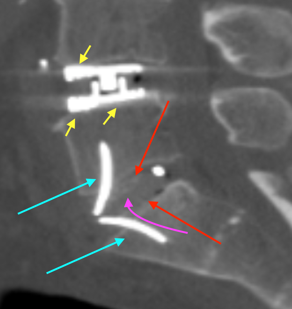
Image 2 (below): A side-on post-op. lumbar CT view of L4/5 and L5/S1. The artificial disc is at the upper disc level (L4/5); the cage is at the lower disc level (L5/S1).
The artificial disc has hydroxyapatite powder-coated roughened titanium outer plate surfaces that are meant to fuse with the patient’s bony endplates (tips of yellow arrows). The polycarbonate urethane (PCU) with silicone gel voids and titanium “monoblock” core of this artificial disc remains mobile, however, for motion-preservation.
The cage (made of superstrong PEEK biocompatible plastic) has large, integrated serrated gull-wing titanium anchors (blue arrows) as well as large serrated surface area contact points with the patient’s bony endplates (tips of red arrows) for surface impaction and fusion. It also has a core (pink arrow-tip region) of “Boost Ultra” (sterile demineralized bone granules/fibres) + blood (sterile patient’s own blood) for internal fusion (a bridge between the adjacent vertebral bodies, which can take up to a year to form and mature).
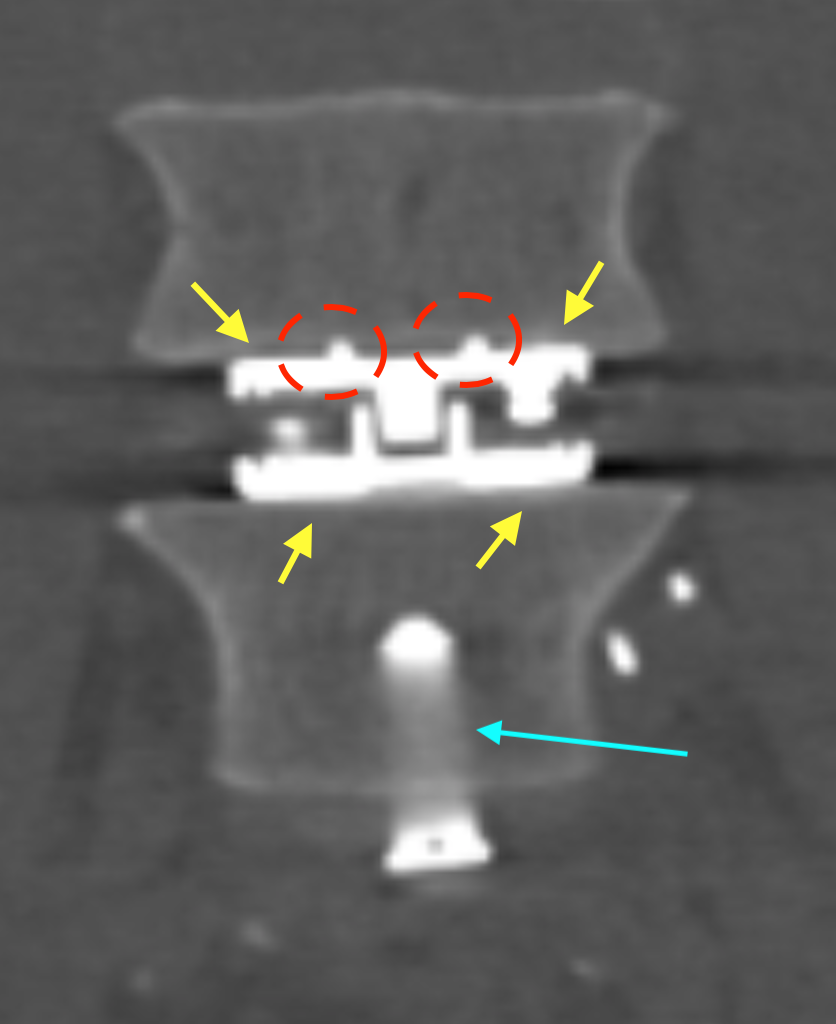
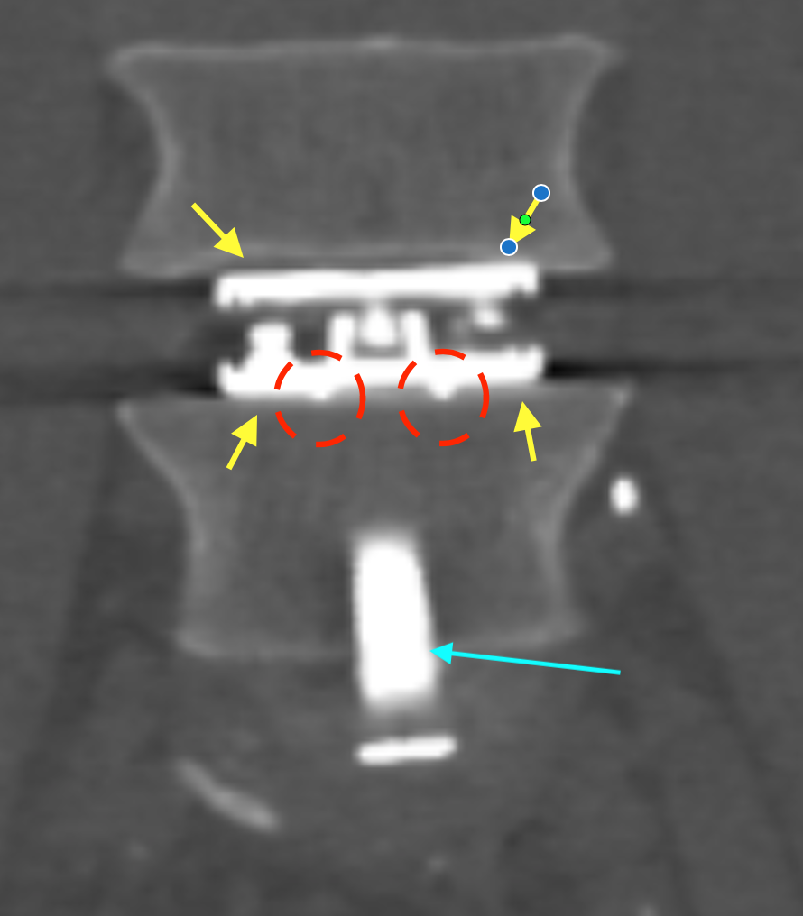
Images 3 & 4 (below): Front-on post-op. lumbar CT views of L4/5 and L5/S1. The artificial disc is at the upper disc level (L4/5); the cage is at the lower disc level (L5/S1).
The artificial disc‘s two titanium plates also have multiple solid spikes (dashed red ovals) that enter into the cartilage and bony endplate surfaces for anchoring / extra ‘purchase’ beyond their usual solid surface contact points (tips of yellow arrows). Parts of the cage‘s titanium gull-wing anchor system are also seen (blue arrows).