Elegant ‘Hybrid’ Technologies for Cervical Reconstruction
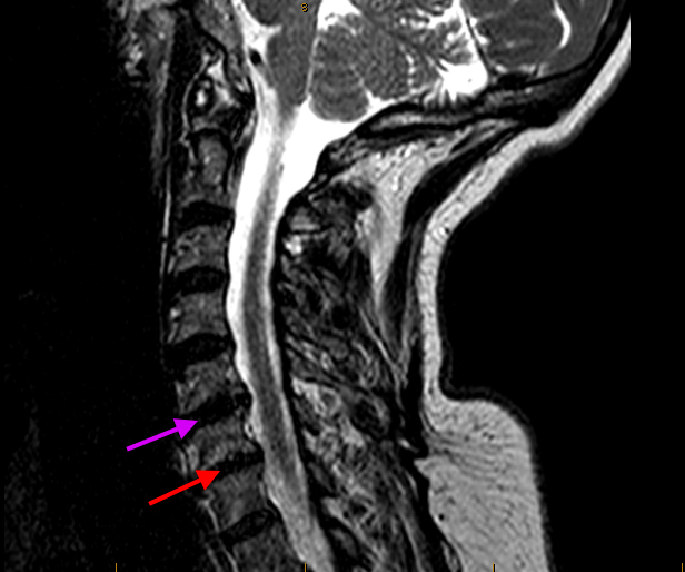
This patient presented with chronic neck and left arm pain. Her pre-operative MRI image below shows bulging degenerative cervical discs at C5/6 (magenta arrow) and C6/7 (red arrow). The C6/7 disc (red arrow) was like a hardened (calcified), deflated ‘car tyre’ affecting the surrounding bone (Modic change) and nerve tissue (stenosis, impingement, radiculopathy).
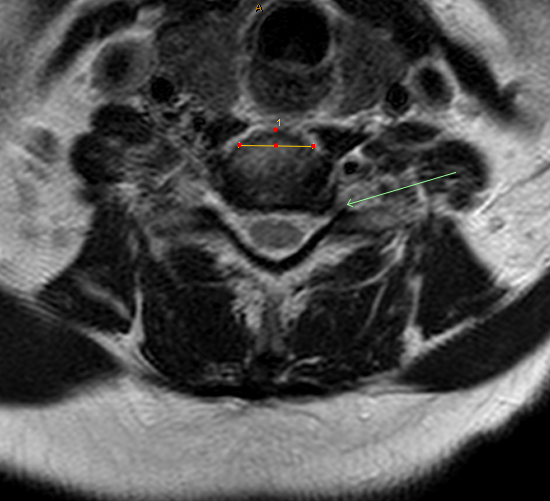
The cross-sectional (axial) MRI image below shows an area of impingement (herniated firm disc-osteophyte) compressing a C7 nerve root (green arrow). The yellow line (numbered ‘1’) shows a pre-operative side-to-side (transverse) measurement to size up the correct custom-shape cage system that will then be introduced into this space once the whole degenerate disc is cleared using minimally invasive surgical techniques.
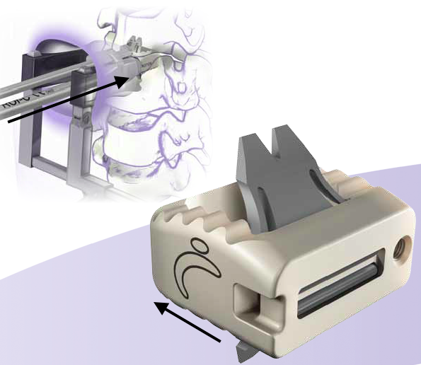
For the very degenerate calcified C6/7 disc space (which in this case was not anatomically suitable for an artificial mobile disc), I chose an elegant Roi-C cage (LDR / Device Technologies; image below). Lordotic, no screws, no plates, and a central bio-active core filled with a bone-fusion promoting compound/matrix (e.g., BoneCrunch, or Genex putty, or iFactor). The titanium ‘gullwing’ anchors will hold the cage in place. The direction of cage placement from the front of the neck is shown by the black arrows. There is minimal disturbance to natural tissues when using an anatomical corridor and retractors to gently spread tissues and access a path between them to the front of the cervical spine. From here, the degenerate disc is carefully sequentially removed using advanced techniques and instruments and steady hands under direct visualisation of the operating microscope and real-time X-ray confirmation.
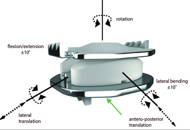
For the less degenerate C5/6 disc space (which was anatomically suitable for an artificial mobile disc), I chose the excellent and time-tested Mobi-C disc (LDR / Device Technologies; image below). The hydroxyapatite+titanium-coated surfaces with ‘teeth’ will hold the disc in place against the cartilaginous ‘endplate’ surfaces. The direction of disc placement from the front of the neck is shown by the green arrow.
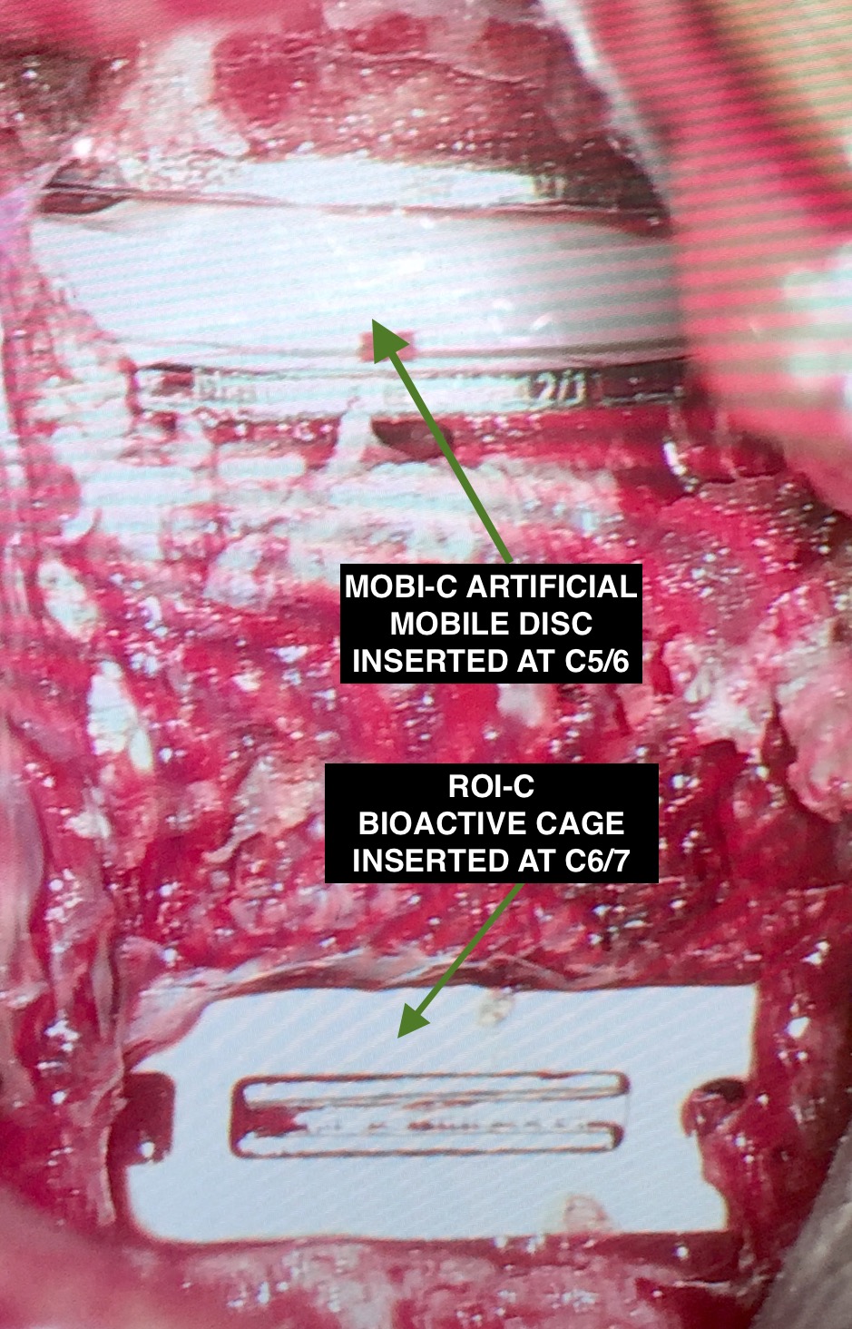
This is what the two prostheses look like in the cervical spine at the end of the operation before I closed up:
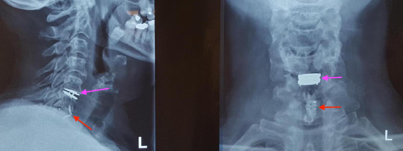
The post-operative X-ray shows the Mobi-C disc (magenta arrow) and Roi-C cage (red arrow), with preservation of cervical lordosis.
The patient awoke from surgery immediately noticing relief of her symptoms, and is making an excellent post-operative recovery having been discharged from hospital after a brief planned stay prior to her interstate travel back home.
Techniques and technologies have come a long way for spinal reconstruction, and when used judiciously, should result in nice outcomes. Please take a moment to look through the other Blogs on this Website, and our patients’ Google Reviews.
< Back to blog