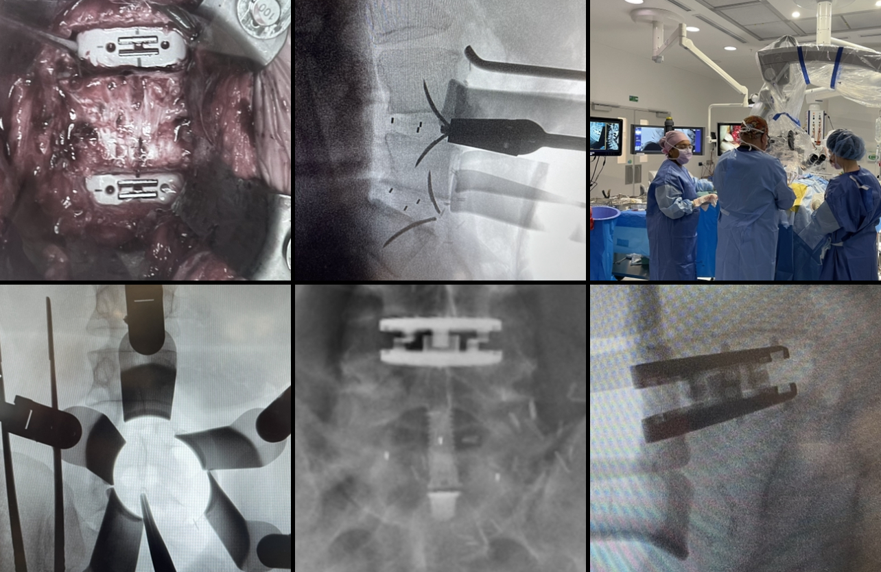
Here are four lovely patient examples of damaged discs in injured or degenerative spines being restored functionally and structurally to help alleviate patients’ pain and disability. I use state-of-the art, unique French prosthetics. The surgery to access the spine in these particular operations is done via the abdomen, a single incision through the skin and an underlying muscle (which is repaired at the end of the procedure), and a vascular surgeon facilitates that part. My bit involves carefully removing each damaged disc, and replacing it with a suitable prosthetic.
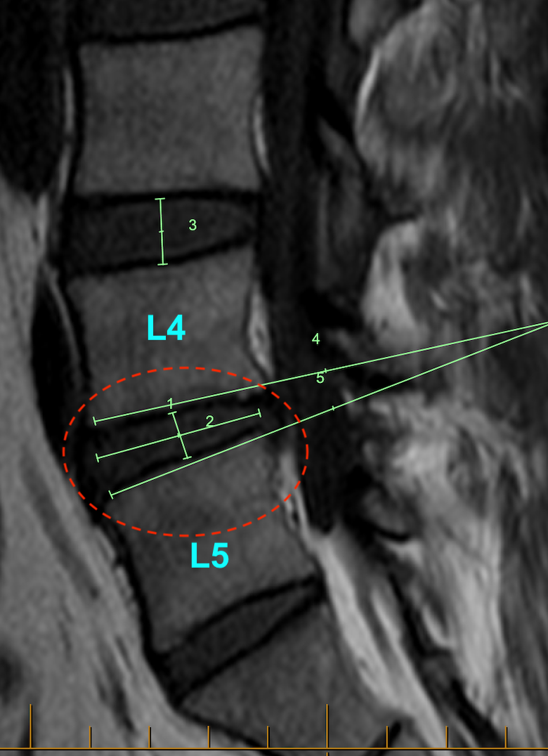
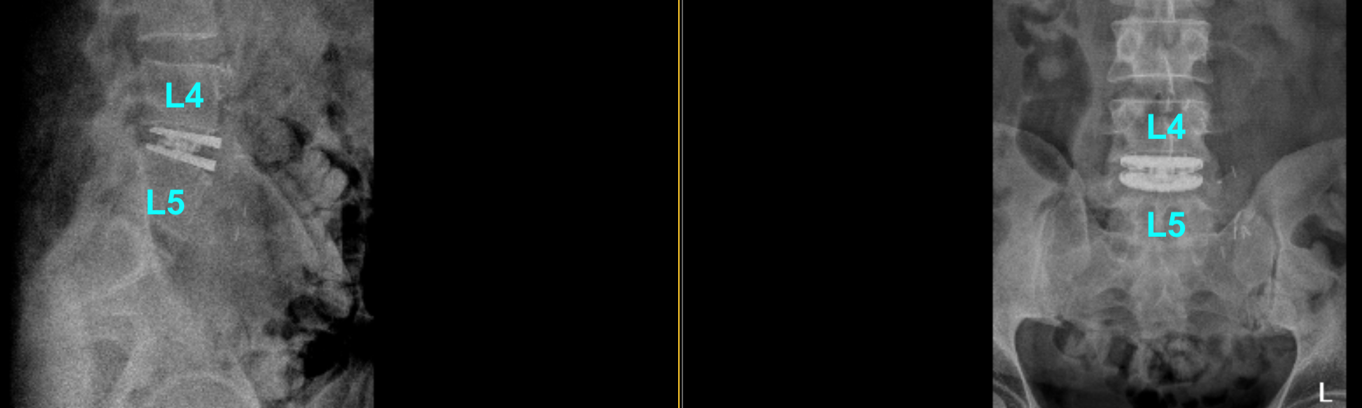
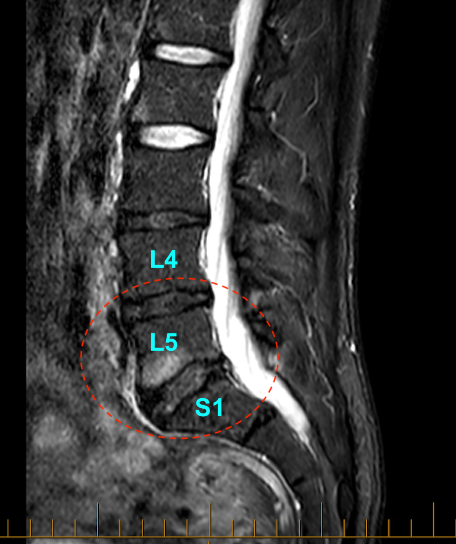
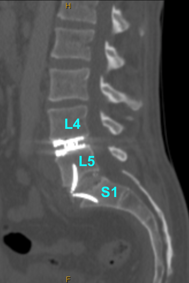
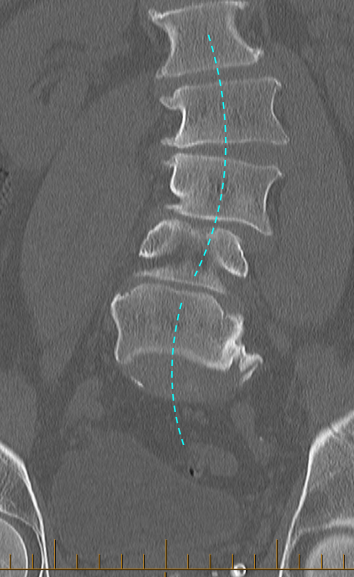
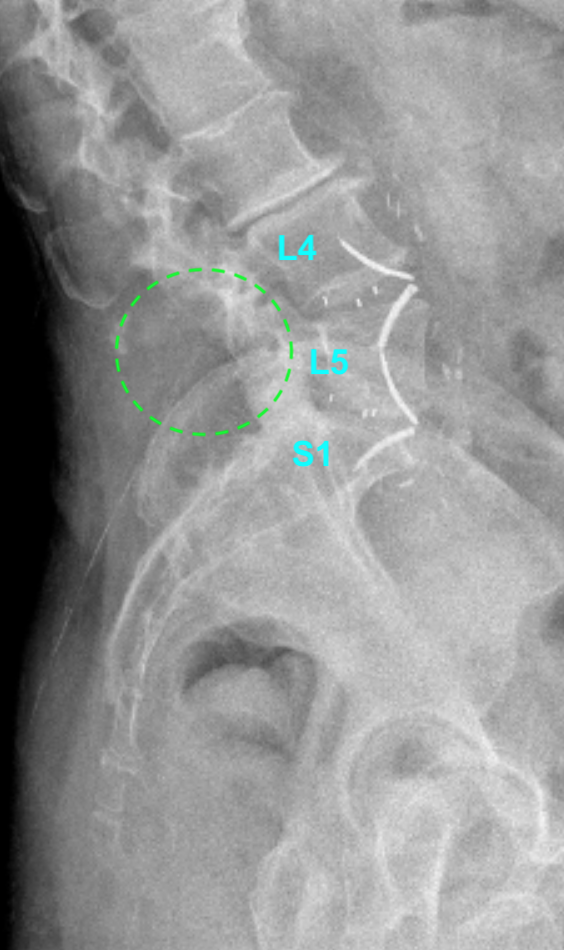
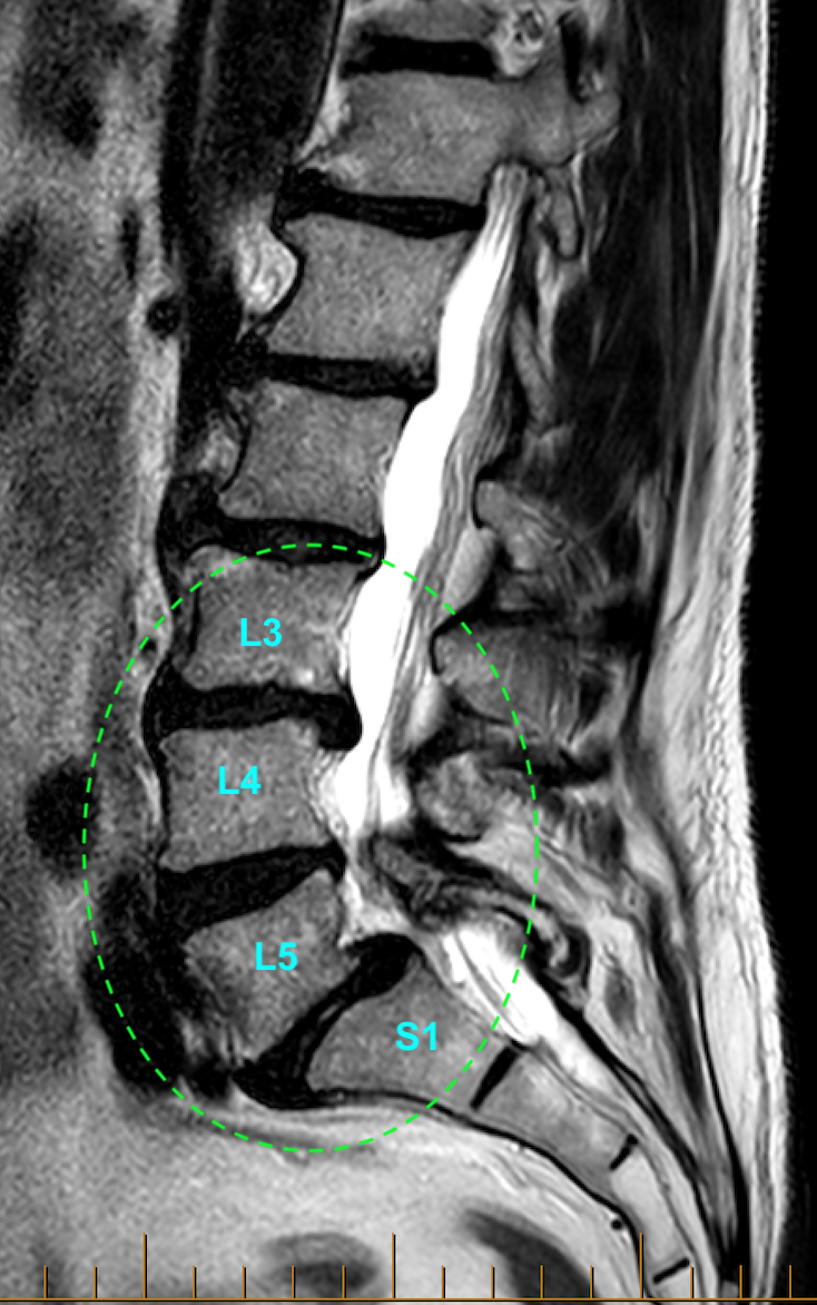
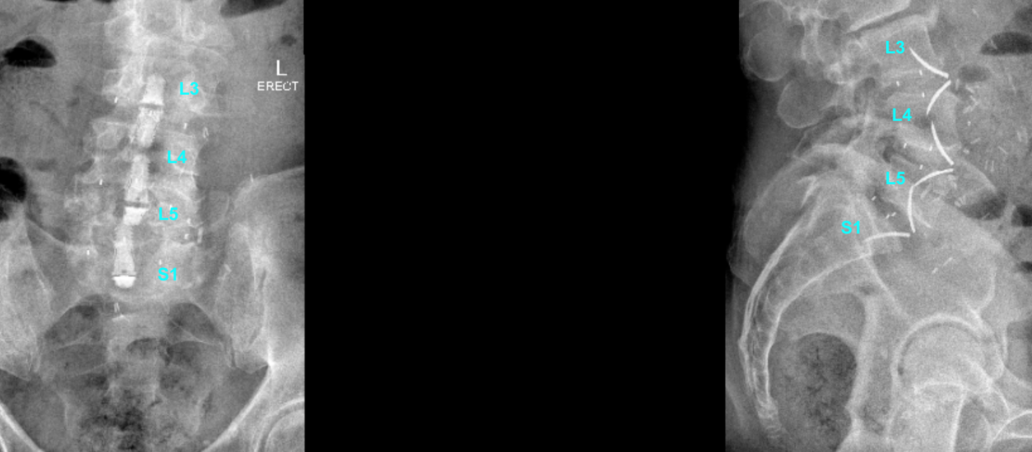
Image 1A (above). This security guard had an injured L4/5 disc, with height and hydration loss (desiccation) and a posterior bulge (herniation) with neural compression.Image 1B (above). The solution for him was a L4-5 artificial disc (total disc arthroplasty/TDA aka total disc replacement/TDR).Image 2A (above). This business owner, an active and athletic chap, wore out his lower lumbar discs, and was quite limited by that.Image 2B (above). The solution for him was a L4-5 artificial disc (total disc arthroplasty/TDA aka total disc replacement/TDR) and a L5-S1 sleek fusion cage. This is also known as a “hybrid” reconstruction. A lovely restoration of disc heights and lumbar lordosis, and indirect neural decompression. Find out more about these prosthetics by CLICKING HERE.Image 3A (above). This lady presented with substantial back pain and leg symptoms from a sagging, scoliotic spine.Image 3B (above). The solution for her was a pair of sleek fusion cages (dual-level anterior lumbar interbody fusion, ALIF) for stability and height restoration, and posterior spinal decompression to free up the compressed nerves directly. That was done in a two-stage operation with real-time sensory and motor nerve electrophysiological monitoring.Image 4A (above). This painter has had a hard working life, and his very degenerate spine shows that. However, at L5/S1 there is a Meyerding Grade 1-2 spondylolisthesis (forward “slip”) that was caused by a birth defect in the L5 pars interarticularis (aka “pars defect”). That resulted in a structural compromise leading to “isthmic spondylolisthesis” and secondary scoliosis. His sagging, collapsing spine needed fixing, and the first stage of that has recently been completed by me (see below).Image 4B (above). The solution for him was three sleek fusion cages (three-level anterior lumbar interbody fusion, ALIF) for stability and height restoration, as stage 1. Stage 2 will be a posterior spinal decompression and L5-S1 Mazor-robot assisted percutaneous (minimally invasive) screws and rods, to free up compressed nerves directly and to “back-up” the fusion cage with the extra instrumentation, owing to his pars defect. Each stage is done with real-time sensory and motor nerve electrophysiological monitoring.Collage of anterior spinal surgery. Prosthetic deployment/implantation (top middle, lower left, and lower right). Top left shows dual ALIF prosthetics immediately after implantation. The single abdominal incision is closed with restoration of the myofascial layer and self-dissolving skin sutures.
The same beautiful prosthetics and work can be used in the cervical spine. CLICK HERE for a lovely example of that.
To learn more about the amazing French prosthetics I use, CLICK HERE. Art meets science meets medicine!